Home
/ Compact Bone Diagram Endosteum : BIOSTR 301 Study Guide (2013-14 Ronald Stenkamp ... : Cc licensed content, specific attribution.
Compact Bone Diagram Endosteum : BIOSTR 301 Study Guide (2013-14 Ronald Stenkamp ... : Cc licensed content, specific attribution.
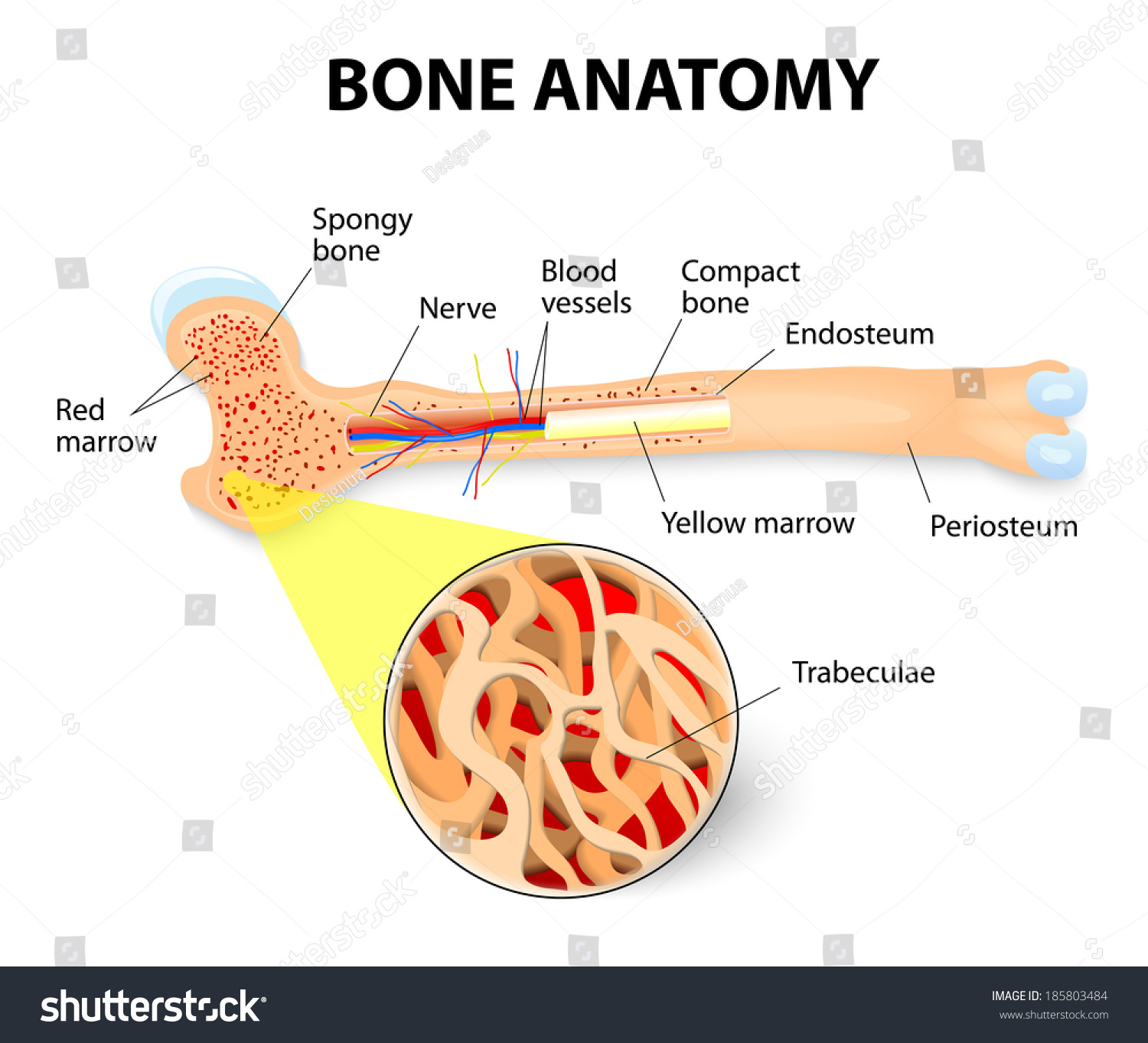
Compact Bone Diagram Endosteum : BIOSTR 301 Study Guide (2013-14 Ronald Stenkamp ... : Cc licensed content, specific attribution.. The densest and strongest bones in the body. It is made up of connective. • the sections are then cut and stained with hx and eosin to demonstrate: Moreover, periosteum and endosteum cover the compact bone from outside and inner surface respectively. Spongy bone, compact bone, articular cartilage, endosteum.
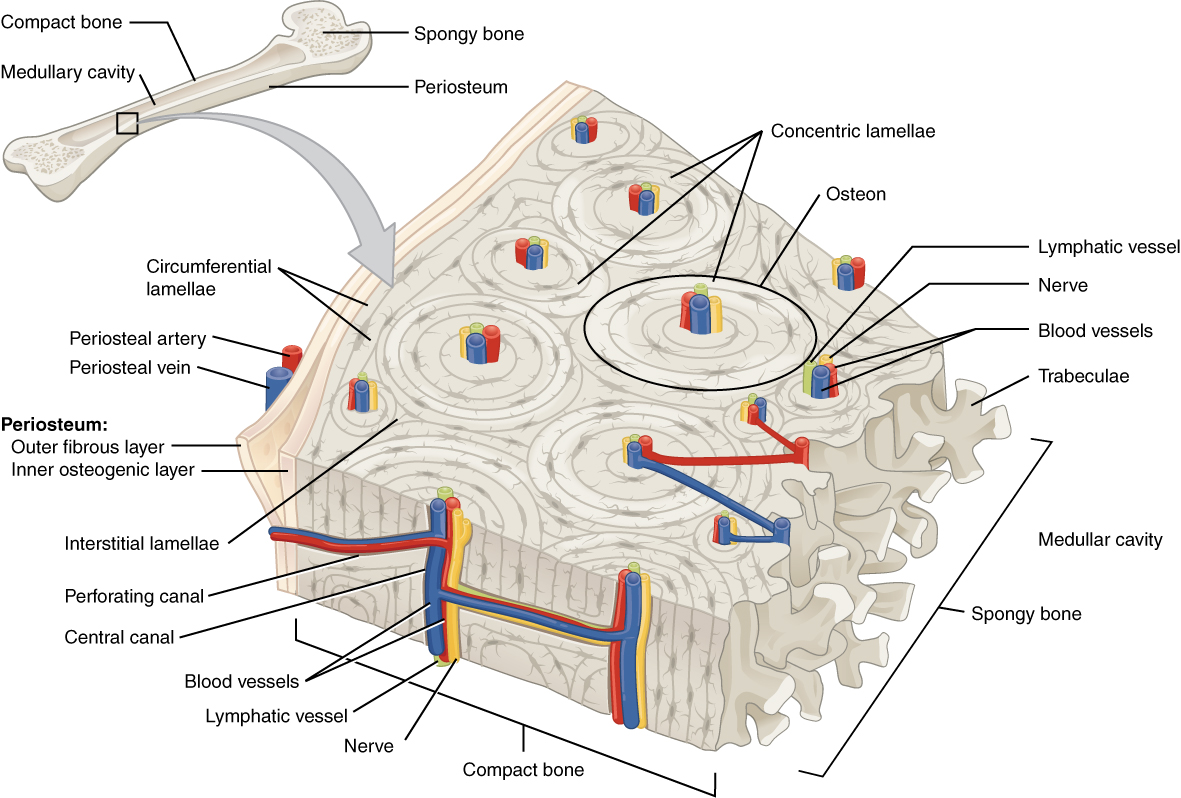
To recognise bone and understand its structure and to understand the processes by which bone can be formed. To know the structures of a synovial joint and a symphysis joint (intervertebral disc). Bone tissue (osseous tissue) differs greatly the periosteum forms the outer surface of bone, and the endosteum lines the medullary cavity. Identify the structures that compose compact and spongy bone. The outer and inner regions contain layers of lamellar bone that run circumferentially around the entire bone.
Anatomy Long Bone Periosteum Endosteum Bone Stock Vector ... from image.shutterstock.com These bones tend to support weight and help movement. Moreover, periosteum and endosteum cover the compact bone from outside and inner surface respectively. Compact bone forms the outer 'shell' of bone. It is a thin covering that surrounds it coats the inner compact bone and the trabeculae of the spongy bone. Endosteum is a thin, soft, connective tissue, lining the cavity of long bones like humerus and femur. A diagram of the anatomy of a bone, showing the compact bone. The endosteum (plural endostea) is a thin vascular membrane of connective tissue that lines the inner surface of the bony tissue that forms the medullary cavity of long bones. The densest and strongest bones in the body.
Compact bone and spongy bone:
The densest and strongest bones in the body. To view the structure of compact bone, scientists. These are mostly compacted bone with little marrow and include most of the bones in the limbs. Compact bone that forms the shafts of long bone consists of two structures. On free bony surfaces of the periosteum and endosteum. Membranes, including the endosteum and periosteum. It is found in bones such as the humerus and the. The inner surface of compact bone is lined by a thin, cellular layer, the endosteum. Definition and functions the endosteum is a structure in the middle of bone tissue and bone marrow. Cancellous bones, compact bone, cortical bone, diaphyses, haversian canal, lamella, marrow cavity, osseous tissue, osteons, spongy bone the inner surface of the bone is covered by the endosteum, a thin, vascular connective tissue, which lines the marrow cavity of the long bones. Compact bone forms the outer 'shell' of bone. Consists of compact bone and the medullary cavity where the bone marrow is stored. Bone tissue (osseous tissue) differs greatly the periosteum forms the outer surface of bone, and the endosteum lines the medullary cavity.
Flat bones, like those of the cranium, consist. This endosteal surface is usually resorbed during long periods of malnutrition, resulting in less cortical thickness. Cc licensed content, specific attribution. • the sections are then cut and stained with hx and eosin to demonstrate: Bone tissue (osseous tissue) differs greatly the periosteum forms the outer surface of bone, and the endosteum lines the medullary cavity.
Archivo:605 Compact Bone.jpg - Wikipedia, la enciclopedia ... from upload.wikimedia.org Osteocytes synthesize bone and reside on the surfaces of bone: Bone tissue (osseous tissue) differs greatly the periosteum forms the outer surface of bone, and the endosteum lines the medullary cavity. It is made up of connective. The outer and inner regions contain layers of lamellar bone that run circumferentially around the entire bone. Spongy bone, compact bone, articular cartilage, endosteum. To know the structures of a synovial joint and a symphysis joint (intervertebral disc). To view the structure of compact bone, scientists. In other terms, they are the cortical bones;
Endosteum is a thin, soft, connective tissue, lining the cavity of long bones like humerus and femur.
Bones are treated with nitric acid to remove their calcium. The endosteum is thin connective. It is found in bones such as the humerus and the. • the sections are then cut and stained with hx and eosin to demonstrate: Osteocytes synthesize bone and reside on the surfaces of bone: Compact bone forms the outer 'shell' of bone. This endosteal surface is usually resorbed during long periods of malnutrition, resulting in less cortical thickness. Bone anatomy marrow cell human long structure diagram spongy body osteoporosis medical vector biology compact matrix blood educational joint osteon system anatomical calcium cartilage disease endosteum. Flat bones, like those of the cranium, consist. Sclerostin inhibits bone formation mostly by antagonizing lrp5/6, thus inhibiting wnt signaling. It is a thin covering that surrounds it coats the inner compact bone and the trabeculae of the spongy bone. To recognise bone and understand its structure and to understand the processes by which bone can be formed. Endosteum is a thin, soft, connective tissue, lining the cavity of long bones like humerus and femur.
Osteocytes synthesize bone and reside on the surfaces of bone: • a compact cortical shaft or diaphysis, (comprising a cylinder of compact bone, its cavity (medulla) being filled with spongy cancellous bone containing bone marrow). Compact bone and spongy bone: Describe how bones are nourished and innervated. The densest and strongest bones in the body.
Figure Periosteum and bone marrow/endosteum give rise to ... from www.researchgate.net Are located in the periosteum and endosteum. Moreover, periosteum and endosteum cover the compact bone from outside and inner surface respectively. Anatomy of the long bone. Spongy bone, compact bone, articular cartilage, endosteum. The endost is covered by internal general plates, enclosing. To view the structure of compact bone, scientists. It is a thin covering that surrounds it coats the inner compact bone and the trabeculae of the spongy bone. Bone anatomy marrow cell human long structure diagram spongy body osteoporosis medical vector biology compact matrix blood educational joint osteon system anatomical calcium cartilage disease endosteum.
The endosteum is thin connective.
A similar layer, the endosteum lines the cavities. To view the structure of compact bone, scientists. Definition and functions the endosteum is a structure in the middle of bone tissue and bone marrow. To know the architecture of compact and spongy (cancellous) bone. Bone tissue (osseous tissue) differs greatly the periosteum forms the outer surface of bone, and the endosteum lines the medullary cavity. Unit 4, test 2 at university of louisiana at monroe. Cancellous bone heals faster than cortical bone because of its vascularization leads. This endosteal surface is usually resorbed during long periods of malnutrition, resulting in less cortical thickness. The outer and inner regions contain layers of lamellar bone that run circumferentially around the entire bone. They are very difficult to distinguish from the surrounding connective tissue cells. Moreover, periosteum and endosteum cover the compact bone from outside and inner surface respectively. Flat bones, like those of the cranium, consist. The densest and strongest bones in the body.
On free bony surfaces of the periosteum and endosteum compact bone diagram. • the sections are then cut and stained with hx and eosin to demonstrate:
